Advanced Example exp_microdosimetry
Responsible Geant4 Collaborators: Giuliana Milluzzo (b), Susanna Guatelli (a) and Francesco Romano (b)
Current external contributors: D. Bolst (a), J. Magini (c), G. Parisi (c)
Past contributors: J. Davis (a), M. G. Pia (d)
(a) Centre For Medical Radiation Physics, Wollongong University, Australia. (b) INFN Sezione Catania, Catania, Italy. (c) University of Surrey, United Kingdom. (d) INFN Sezione di Genova, Genova, Italy.
Short description
The exp_microdosimetry example, originally named Radioprotection, models different detectors for microdosimetry for radiation protection in space applications. The example lets the user choose between the models of a simplified diamond (1), a micro-diamond (2) and a silicon microdosimeter (3):
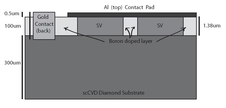
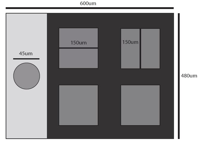
1) A sшmplified diamond microdosimeter (in Figure 1 and 2), based on the detector developed by Prof. Anatoly Rosenfeld and his team at the Centre For Medical Radiation Physics, CMRP, University of Wollongong, NSW, Australia. The design of the device is documented in J. Davis, et al., “Characterisation of a novel diamond-based microdosimeter prototype for radioprotection applications in space environments”, IEEE Transactions on Nuclear Science, Vol. 59, pp. 3110-3116, 2012.
2) A microdiamond detector, based on the detectors developed by the Research Group of The University of Rome “Tor Vergata”. The design and performances of the detector are documented in C. Verona et al., “Spectroscopic properties and radiation damage investigation of a diamond based Schottky diode for ion-beam therapy microdosimetry”, Journal of Applied Physics, vol. 118, 2015, and in C. Verona et al., “Toward the use of single crystal diamond based detector for ion-beam therapy microdosimetry”, Radiation Measurements, vol. 110, 2018.
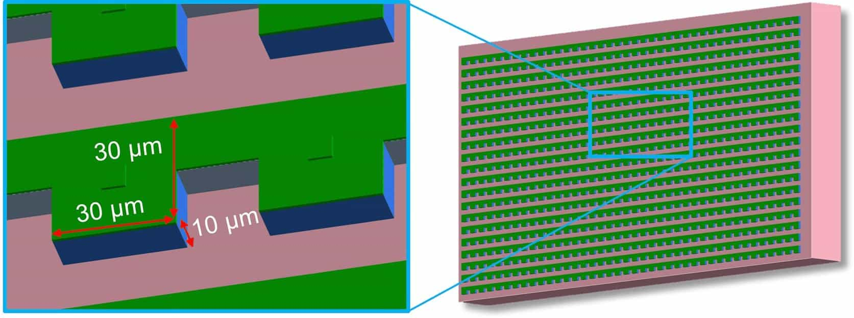
3) A silicon microdosimeter (in Figure 3), based on the “Bridge” microdosimeter, developed by the Centre For Medical Radiation physics, University of Wollongong, documented in chapter 7 of the PhD Thesis of D. Bolst “Silicon microdosimetry in hadron therapy using Geant4”, available online at https://ro.uow.edu.au/theses1/619/
All the microdosimeters are set in vacuum and it is possible to select among the avilable detectors via a macro command (/geometrySetup/selectDetector “…”), which is included in the “geometry.mac” macro.
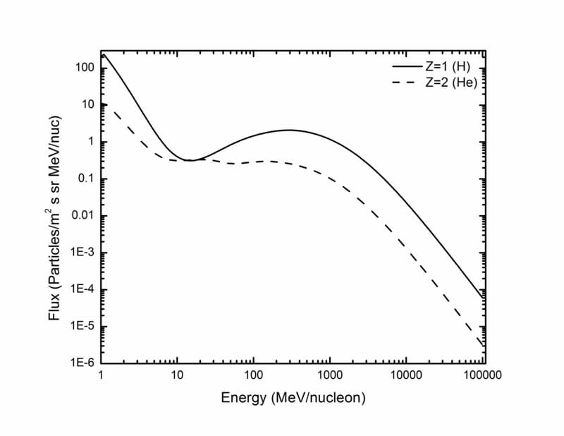
An isotropic field of Galactic Cosmic Rays (GCR) protons or alpha particles is incident on the device. The particle spectra are shown in Figure 4 and have been obtained from SPENVIS. They are based on CREME96 (A J. Tylka, et al. IEEE Trans. Nucl. Sci., Vol. 44 (6), 1997). The energy deposition is calculated in the sensitive detectors.
This example shows how to:
- model a realistic isotropic field of GCRs by means of the General Particle Source;
- model a realistic detector in Geant4;
- retrieve the information of secondary particles that originated in the sensitive volume of the detector.
- calculate the energy deposition in the sensitive volumes of a detector. Please refer to the README file to have more detailed information on how to run the example.
Last updated: 23/02/2022 by S. Guatelli